
CardaxEP
Simulate cardiac rhythms, pacing, mapping, and ablation in a four-chamber heart.
macOS 14 or later
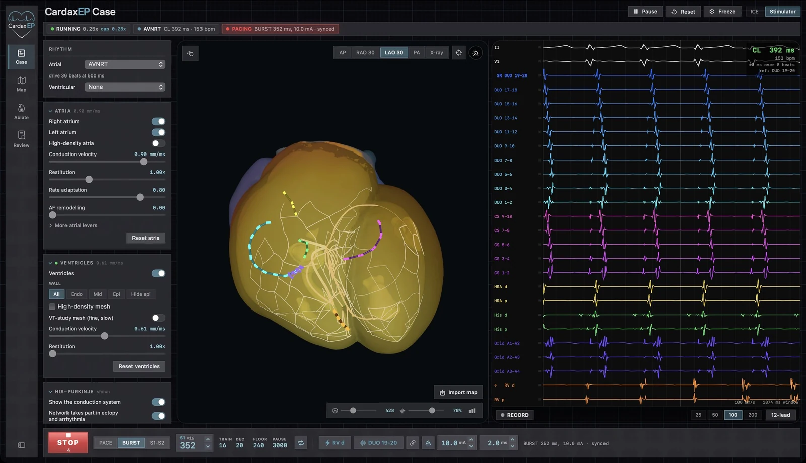
Four-chamber electrical model
Atrial tissue, ventricular muscle, and the conduction system participate in the same simulated beat. The wavefront, catheter recordings, and surface leads all update as the tissue responds.
- Follow anisotropic activation through the biatrial model or through a volumetric ventricular mesh with fiber orientation and differences across the wall.
- Inspect the dual-pathway AV node and His–Purkinje network, including forward and reverse conduction.
- Switch between anatomical, fluoroscopy-style, and activation views to relate a signal to where it started and how it traveled.
Rhythm and induction library
Start with baseline conduction, then compare focal, reentrant, and fibrillatory rhythms under controlled conditions.
- Explore sinus and junctional rhythms, focal atrial tachycardia, typical and reverse typical flutter, scar-related atypical flutter, and atrial fibrillation.
- Compare AV nodal reentrant tachycardia (AVNRT) with atrioventricular reentrant tachycardia (AVRT) and see how each responds to pacing.
- Select anatomically located premature ventricular contractions (PVCs), test a scar substrate for ventricular tachycardia (VT), and examine ventricular fibrillation (VF).
- Use an unknown case to interpret the signals and build a map before revealing the rhythm.
Ventricular ectopy and wall depth
Move the starting point of a premature ventricular beat and compare how that change appears in the activation map and the 12-lead ECG.
- Select named origins in the outflow tracts, septum, ventricular walls, or conduction system where the model supports them.
- Set the focus at the endocardial, mid-myocardial, or epicardial depth where anatomy permits.
- Compare a single PVC with couplets, bigeminy, trigeminy, or a specified ectopic burden and coupling interval.
Catheters, electrograms, and 12-lead ECG
Follow local and far-field signals alongside the simulation. Electrograms and surface leads are computed from the tissue model as it runs.
- Position and display simulated multipolar, coronary sinus, His, right ventricular, and high-density mapping catheters.
- Inspect unipolar and bipolar channels and change the sweep, lead selection, or arrangement of the recording.
- Open a simulated 12-lead ECG and compare intervals, axis, and morphology as you move the origin of an ectopic beat or alter conduction.
Programmed stimulation and entrainment
Use the stimulator to test the mechanism of a rhythm by changing where and when a stimulus arrives.
- Run a drive train, add timed S1–S2 extrastimuli, or deliver a burst from a selected catheter electrode.
- Adjust cycle length and coupling intervals, then inspect capture, conduction, and the response after pacing stops.
- Compare maneuvers across pacing sites and record the response for review with calipers.
Electroanatomic mapping
Build activation and voltage maps from acquired catheter electrograms. Use the live activation display as a separate view of the simulated tissue.
- Choose a reference channel and a window of interest; acquire the current beat or collect accepted beats continuously.
- Move the mapping catheter across the selected surface and compare local activation timing and voltage.
- Review acquired points, discard the most recent beat, or save a map. The map import workspace can bring supported map captures into a substrate study.
Substrate and simulated ablation
Change the properties of a model and test whether a circuit survives. The resulting effects come from the simulated wavefront meeting the new tissue conditions.
- Vary conduction and refractoriness or compare normal and remodeled tissue settings.
- Draw regions of scar and slow conduction and control how deep the change extends into the ventricular wall.
- Place a point lesion or a line across a suspected pathway, then look for slowing, block, reorganization, or termination.
Study recording and review
Capture an induction, pacing maneuver, or rhythm transition and revisit it without rerunning the case.
- Record surface and intracardiac channels and inspect the corresponding pacing log.
- Pan through the saved tracings, change the visible time window, and move paired calipers while keeping their measured interval fixed.
- Save the study or a standalone map to compare runs or discuss findings with a learner.
Integrated intracardiac echo
The desktop lab can display synthetic intracardiac echocardiography (ICE) alongside the map and recordings.
- Use the simulated catheter and standard-view presets to see how orientation affects the ultrasound plane.
- Compare the image with the anatomical orientation view. Visit the dedicated ICE page for catheter and transseptal training capabilities.
In active development · Education and research simulation
Ventricular tachycardia transitions to fibrillation
Follow the ventricular activation map and simulated ECG together as a regular ventricular rhythm breaks into disorganized activity.
Open videoAVNRT: right ventricular overdrive pacing
Watch a pacing maneuver in the complete CardaxEP workspace with the surface ECG, coronary sinus, His, mapping, and right ventricular channels visible.
Open videoAVRT: right ventricular overdrive pacing
Compare the tracing during a paced maneuver in simulated AV reentry with the AVNRT example.
Open videoTypical atrial flutter
Orbit the biatrial model to follow the organized circuit and see how it relates to the surrounding anatomy.
Open videoAtypical atrial flutter around scar
Follow activation bars on a simulated atrial substrate with scar, and compare the wavefront with the typical flutter example.
Open videoAtrial fibrillation
The camera circles the high-density atrial model while changing fronts propagate across the tissue.
Open videoScar-related ventricular tachycardia
Watch activation bars pass through a narrow conducting channel in a drawn ventricular scar.
Open videoPVC activation map: RV outflow tract septum
Find the earliest activation in red and follow the color progression to the latest activation in purple.
Open videoConduction system in sinus rhythm
A translucent four-chamber view shows the His–Purkinje network through a simulated sinus beat.
Open videoWhole-heart activation in sinus rhythm
Follow one beat through the chambers as activation bars move over the surface of the model.
Open videoAV nodal reentry and electrograms
A close view pairs the rotating four-chamber heart with the scrolling surface ECG and intracardiac tracings.
Open video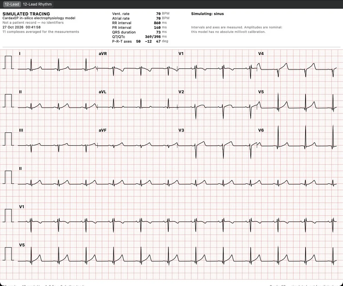
Computed 12-lead ECG
A sinus-rhythm tracing generated from the simulated tissue with rate, intervals, and axes displayed.
Open full image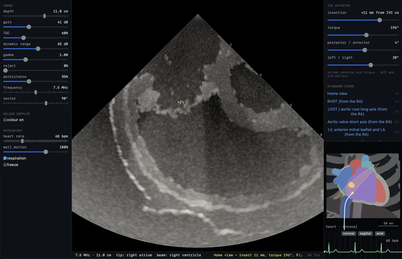
Intracardiac echo in the desktop lab
See the synthetic ultrasound plane alongside catheter steering controls, standard views, and an anatomy inset.
Open full image